Definition of breast cancer: Cancer that forms in tissues of the breast. The most common type of breast cancer is ductal carcinoma, which begins in the lining of the milk ducts (thin tubes that carry milk from the lobules of the breast to the nipple). Another type of breast cancer is lobular carcinoma, which begins in the lobules (milk glands) of the breast. Invasive breast cancer is breast cancer that has spread from where it began in the breast ducts or lobules to surrounding normal tissue. Breast cancer occurs in both men and women, although male breast cancer is rare.
Estimated new cases and deaths from breast cancer in the United States in 2013:
New cases: 232,340 (female); 2,240 (male)
Deaths: 39,620 (female); 410 (male)
The Breasts
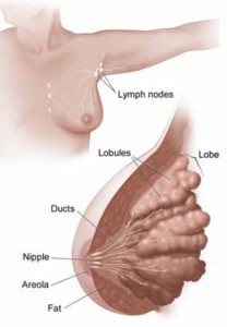
Inside a woman’s breast are 15 to 20 sections (lobes). Each lobe is made of many smaller sections (lobules). Lobules have groups of tiny glands that can make milk.
After a baby is born, breast milk flows from the lobules through thin tubes (ducts) to the nipple. Fibrous tissue and fat fill the spaces between the lobules and ducts.
This picture shows the lobes and ducts inside the breast. It also shows lymph nodes near the breast.
Cancer Cells
Cancer begins in cells, the building blocks that make up all tissues and organs of the body, including the breast.
Normal cells in the breast and other parts of the body grow and divide to form new cells as they are needed. When normal cells grow old or get damaged, they die, and new cells take their place.
Sometimes, this process goes wrong. New cells form when the body doesn’t need them, and old or damaged cells don’t die as they should. The buildup of extra cells often forms a mass of tissue called a lump, growth, or tumor.
Tumors in the breast can be benign (not cancer) or malignant (cancer):
Benign tumors:
- Are usually not harmful
- Rarely invade the tissues around them
- Don’t spread to other parts of the body
- Can be removed and usually don’t grow back
Malignant tumors:
- May be a threat to life
- Can invade nearby organs and tissues (such as the chest wall)
- Can spread to other parts of the body
- Often can be removed but sometimes grow back
Breast cancer cells can spread by breaking away from a breast tumor. They can travel through blood vessels or lymph vessels to reach other parts of the body. After spreading, cancer cells may attach to other tissues and grow to form new tumors that may damage those tissues.
For example, breast cancer cells may spread first to nearby lymph nodes. Groups of lymph nodes are near the breast under the arm (axilla), above the collarbone, and in the chest behind the breastbone.
When breast cancer spreads from its original place to another part of the body, the new tumor has the same kind of abnormal cells and the same name as the primary (original) tumor. For example, if breast cancer spreads to a lung, the cancer cells in the lung are actually breast cancer cells. The disease is metastatic breast cancer, not lung cancer. For that reason, it’s treated as breast cancer, not lung cancer.
Types
Breast cancer is the most common type of cancer among women in the United States (other than skin cancer). In 2013, more than 232,000 American women will be diagnosed with breast cancer.
The most common type of breast cancer is ductal carcinoma. This cancer begins in cells that line a breast duct. See the picture of the breast ducts. About 7 of every 10 women with breast cancer have ductal carcinoma.
The second most common type of breast cancer is lobular carcinoma. This cancer begins in a lobule of the breast. See the picture of lobules. About 1 of every 10 women with breast cancer has lobular carcinoma.
Other women have a mixture of ductal and lobular type or they have a less common type of breast cancer.
Tests
After you find out that you have breast cancer, you may need other tests to help choose the best treatment for you.
Lab Tests with Breast Tissue
The breast tissue that was removed during your biopsy can be used in special lab tests:
- Hormone receptor tests: Some breast cancers need hormones to grow. These cancers have hormone receptors for the hormones estrogen, progesterone, or both. If the hormone receptor tests show that the breast cancer has these receptors, then hormone therapy is often recommended as part of the treatment plan.
- HER2 test: Some breast cancers have large amounts of a protein called HER2, which helps them to grow. The HER2 test shows whether a woman’s breast cancer has a large amount of HER2. If so, then targeted therapy against HER2 may be a treatment option.
It may take several weeks to get the results of these tests. The test results help your doctor decide which cancer treatments may be options for you.
Triple-negative breast cancer
About 15 of every 100 American women with breast cancer have triple-negative breast cancer. These women have breast cancer cells that…
- Do not have estrogen receptors (estrogen negative)
- Do not have progesterone receptors (progesterone negative)
- Do not have a large amount of HER2 (HER2 negative)
Staging Tests
Staging tests can show whether cancer cells have spread to other parts of the body.
When breast cancer spreads, cancer cells are often found in the underarm lymph nodes (axillary lymph nodes). Breast cancer cells can spread from the breast to almost any other part of the body, such as the lungs, liver, bones, or brain.
Your doctor needs to learn the stage (extent) of the breast cancer to help you choose the best treatment. Staging tests may include…
- Lymph node biopsy: If cancer cells are found in a lymph node, then cancer may have spread to other lymph nodes and other places in the body. Surgeons use a method called sentinel lymph node biopsy to remove the lymph node most likely to have breast cancer cells. The NCI fact sheet Sentinal Lymph Node Biopsy has more information, including pictures of the method.If cancer cells are not found in the sentinel node, the woman may be able to avoid having more lymph nodes removed. The method of removing more lymph nodes to check for cancer cells is called axillary dissection.
- CT scan: An x-ray machine linked to a computer takes a series of detailed pictures of your chest or abdomen. You may receive contrast material by mouth and by injection into a blood vessel in your arm or hand. The contrast material makes abnormal areas easier to see. The pictures from a CT scan can show cancer that has spread to the lungs or liver.
- MRI: A strong magnet linked to a computer is used to make detailed pictures of your chest, abdomen, or brain. An MRI can show whether cancer has spread to these areas. Sometimes contrast material makes abnormal areas show up more clearly on the picture.
- Bone scan: The doctor injects a small amount of a radioactive substance into a blood vessel. It travels through the bloodstream and collects in the bones. A machine called a scanner detects and measures the radiation. The scanner makes pictures of the bones. Because higher amounts of the substance collect in areas where there is cancer, the pictures can show cancer that has spread to the bones.
- PET scan: You’ll receive an injection of a small amount of radioactive sugar. The radioactive sugar gives off signals that the PET scanner picks up. The PET scanner makes a picture of the places in your body where the sugar is being taken up. Cancer cells show up brighter in the picture because they take up sugar faster than normal cells do. A PET scan can show cancer that has spread to other parts of the body.
Questions you may want to ask your doctor about tests
- What did the hormone receptor test show?
- What did the HER2 test show?
- May I have a copy of the report from the pathologist?
- Do any lymph nodes show signs of cancer?
- What is the stage of the disease?
- Has the cancer spread?
- Would genetic testing be helpful to me or my family?
Stages
The stage of breast cancer depends on the size of the breast tumor and whether it has spread to lymph nodes or other parts of the body.
Doctors describe the stages of breast cancer using the Roman numerals 0, I, II, III, and IV and the letters A, B, and C.
A cancer that is Stage I is early-stage breast cancer, and a cancer that is Stage IV is advanced cancer that has spread to other parts of the body, such as the liver.
The stage often is not known until after surgery to remove the tumor in the breast and one or more underarm lymph nodes.
Stage 0
Stage 0 is carcinoma in situ. In ductal carcinoma in situ (DCIS), abnormal cells are in the lining of a breast duct, but the abnormal cells have not invaded nearby breast tissue or spread outside the duct.
Stage IA
The breast tumor is no more than 2 centimeters (no more than 3/4 of an inch) across. Cancer has not spread to the lymph nodes.
© 2007 Terese Winslow. U.S. Govt has certain rights
A tumor that is 2 centimeters is about the size of a peanut, and a tumor that is 5 centimeters is about the size of a lime.
Stage IB
The tumor is no more than 2 centimeters across. Cancer cells are found in lymph nodes.
Stage IIA
The tumor is no more than 2 centimeters across, and the cancer has spread to underarm lymph nodes.
Or, the tumor is between 2 and 5 centimeters (between 3/4 of an inch and 2 inches) across, but the cancer hasn’t spread to underarm lymph nodes.
Stage IIB
The tumor is between 2 and 5 centimeters across, and the cancer has spread to underarm lymph nodes.
Or, the tumor is larger than 5 centimeters across, but the cancer hasn’t spread to underarm lymph nodes.
Stage IIIA
The breast tumor is no more than 5 centimeters across, and the cancer has spread to underarm lymph nodes that are attached to each other or nearby tissue. Or, the cancer may have spread to lymph nodes behind the breastbone.
Or, the tumor is more than 5 centimeters across. The cancer has spread to underarm lymph nodes that may be attached to each other or nearby tissue. Or, the cancer may have spread to lymph nodes behind the breastbone but not spread to underarm lymph nodes.
Stage IIIB
The breast tumor can be any size, and it has grown into the chest wall or the skin of the breast. The breast may be swollen or the breast skin may have lumps.
The cancer may have spread to underarm lymph nodes, and these lymph nodes may be attached to each other or nearby tissue. Or, the cancer may have spread to lymph nodes behind the breastbone.
Stage IIIC
The breast cancer can be any size, and it has spread to lymph nodes behind the breastbone and under the arm. Or, the cancer has spread to lymph nodes above or below the collarbone.
Stage IV
The tumor can be any size, and cancer cells have spread to other parts of the body, such as the lungs, liver, bones, or brain.
Inflammatory Breast Cancer
- Inflammatory breast cancer is a rare type of breast cancer. It occurs in about 1 of every 100 American women with invasive breast cancer.
- The breast looks red and swollen because cancer cells block the lymph vessels in the skin of the breast.
- When a doctor diagnoses inflammatory breast cancer, it’s at least Stage IIIB, but it could be more advanced.


